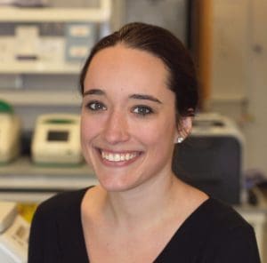
Colors matter to fifth-year Johns Hopkins Ph.D. student Sarah Hadyniak. And what especially matters to her is the order in which they develop—and why that’s potentially important to treat health disorders affecting vision.
Humans have trichromatic color vision, which comes from the presence of three types of cone cells in the eye’s retina. These cone cells are responsible for color perception and react to light of blue, green, and red wavelengths.
Hadyniak’s work has focused on the development and patterning of our blue, green, and red cone cells. Up until recently, we haven’t really understood why or how the different cone colors develop and in what order, she explains.
Green and red cones are very similar in everything with the exception of a protein called opsin, which is responsible for absorbing light in the eye. Hadyniak’s project has been to determine which cone cells develop in the retina first, and the factors involved in the generation of those cone cells. She is part of the Bob Johnston lab.
Because retinal development happens in utero, the lab uses retinal organoids, derived from stem cells and grown in the lab, to study this process. And what Hadyniak has found is that Vitamin A-derived retinoic acid is responsible for the generation of red cones by expression of the red opsin protein. During fetal development, blue cones develop first. After that come green cones, and finally, red.
“We are trying to visualize the generation of these cell types and to look at different transcripts and molecules using a variety of techniques,” she says. Those include RNA sequencing to examine the opsin RNA molecules that differ among cone cells, and in-situ hybridization, which allows her to view RNA molecules within the cell.
Long-range vision
Understanding how the retina’s cone cells develop can also help and treat various diseases, Hadyniak says.
For example, cone development takes place on the X chromosome, she explains. About 10 percent of men are colorblind, but only about .03 percent of women are. The reasoning behind colorblindness has a lot to do with these DNA sequences on the X chromosome.
But beyond colorblindness, her work has the potential to help with macular degeneration: the loss of cone cells as we age. Others in the Johnston lab are collaborating to find ways to transplant retinal stem cells into animal model systems, with a future goal of using them to fight blindness caused by this and other conditions.
In the rotation
All students in the Ph.D. program undergo four two-month rotations during their first year. Hadyniak’s rotations included examining sex determination in the drosophila gonad in Mark Van Doren, Ph.D.’s lab; molecular cloning in zebrafish with Marnie Halpern at the Carnegie Institution for Science – Department of Embryology; and Xin Chen’s lab on tracking asymmetric histone segregation in C. elegans.
Johnston’s lab, Hadyniak says, has an environment that’s fun and full of energy. “Bob loves what he does, and he loves to talk and think about science with everyone. I would pick this lab all over again,” she says.
As a biology major at Boston University, she knew she wanted a Ph.D. When she began researching developmental biology programs, she liked that Hopkins offered a strong umbrella program and emphasized broad knowledge.
For example, all incoming Ph.D. students in it must complete a “Computational Boot Camp,” which is a one-week class to teach students coding languages such as Python and new ways to view and analyze data. “I had never done it before, but it’s given me the fundamentals to use what I’ve learned in more depth,” she says.
Hadyniak enjoys a virtual trade route of information throughout Hopkins. She also gets to work with the James Taylor lab at Hopkins for assistance with some of the RNA data she collects. She collaborates closely with Hopkins’ medical school for tissue samples and organoids. Also she has a solid relationship with the Neitz Lab at the University of Washington, which focuses on genetic tests and treatments for vision disorders; they have graciously provided DNA samples and red and green cone cell ratios to help further her work.
“People at Hopkins are just really friendly and really collaborative. Doors are open, literally and figuratively. People make themselves available to talk to you. And I like the feel of Baltimore, I love the community vibe, I feel like people care a lot about this city,” she adds.
She advises potential students considering any graduate biology program to find a balance, no matter where you go. While it’s crucial to find a lab and a mentor who will support your work, you’ll also need to seek out life beyond the lab.
“Find hobbies and things outside of school and science to keep you going,” she urges. “You will have highs where you’ll want to be in the lab all the time, and then you will have some lows, and you will want a support system, whether it’s friends, family, or hobbies.”
What’s next?
Hadyniak is currently writing her thesis, which will center on the cone receptor patterning. Ideally, she’d like to find a postdoc program once she receives her degree, and stay in academia while doing research. She also has what she describes as a side project, which is trying to map the human retina.
“No one has been able to visualize these cell types before in fixed tissue,” she explains.
Hopkins supports its students in many ways, she says. They send students to conferences, bring in speakers, and discuss other related research in the field. Also, every student must provide regular 30-minute talks about their work within the department, which provides the opportunity to improve one’s scientific presentation skills.
“The whole community is driven to publish and be around other labs doing the same thing. But it helps you in producing quality work,” she says.
